
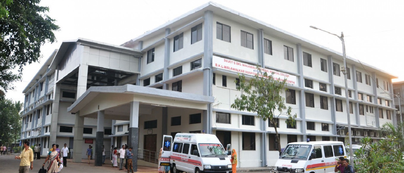
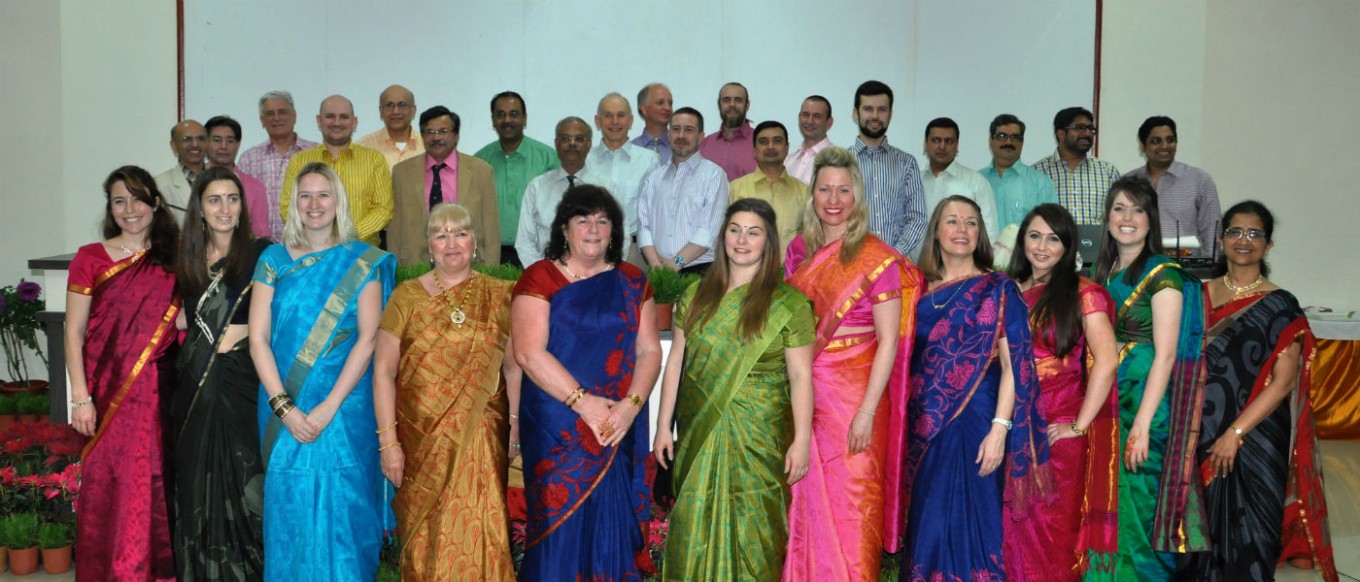
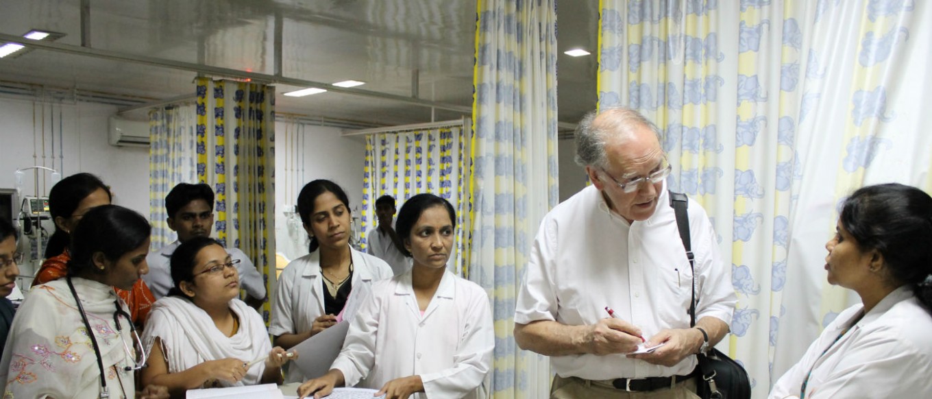


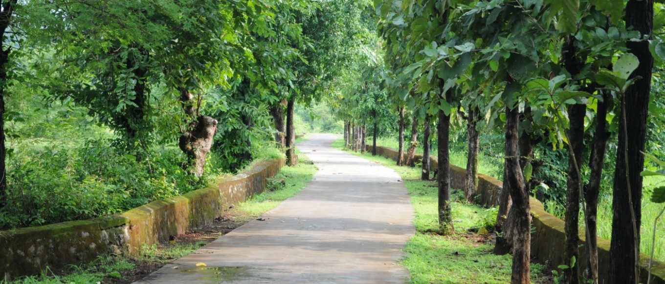
Dervan village in Konkan is situated in one of the most backward regions of Maharashtra State. In 1977, Revered Shri Vithalrao Joshi alias Shree Digambardas Maharaj established Shri Vithalrao Joshi Charities Trust. He had established the Shree Sant Sitarambuwa Walawalkar Charitable Trust in 1969. These two trusts together brought about a total socio-economic transformation of the village…. Read More
- Home
- Specialty Services
- Departments
- Facilities
- Community Service
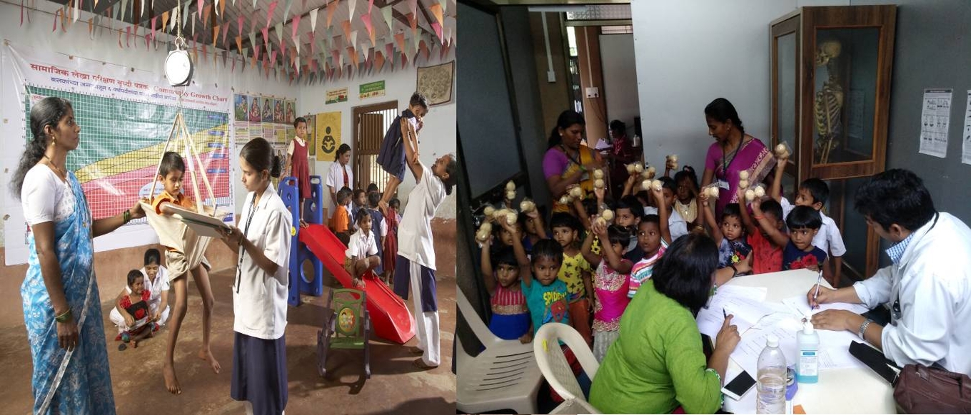
- Walawalkar Laddoo-Gopal Yojana
- Walawalkar Dant Chikitsa Yojana
- Walawalkar Sudama Yojana
- Walawalkar Yashoda Yojana
- Walawalkar Sukanya Yojana
- MOBILE MEDICAL UNIT (NRHM)
- Social & Cultural Activities
- Rural Empowerment and Community Health (REACH)
- Rural Health Training Center (RHTC)
- Urban Health Training Center (UHTC)
- Research
- Education
- Gallery
- Events
- CASE REPORTS
- Donations